Enhancing Precision and Comfort in Root Canal Care with 3-D Imaging Technology
by Dr. Jacqueline S. Allen | Dec 3, 2025 | Cone Beam Computed Tomography

At Phoenix Endodontic Group, we believe that preserving your natural teeth and ensuring your comfort should go hand in hand. That’s why we’ve integrated advanced diagnostic tools—most notably cone-beam computed tomography (CBCT)—into our daily care. This powerful 3-D imaging technology allows our specialists to visualize the smallest details of your tooth structure and surrounding anatomy, leading to more accurate diagnoses, safer procedures, and better long-term outcomes.
Why 3-D Imaging Matters in Endodontic Care
Traditional 2-D dental x-rays can sometimes miss complex or hidden features within a tooth. CBCT, however, provides a comprehensive 3-D view of your tooth, its roots, and nearby bone structures, offering unparalleled diagnostic insight.
With this technology, our endodontists can:
-
Identify root canal anatomy, curvature, and tiny accessory canals.
-
Detect hidden infections, cracks, or resorption that might not appear on traditional films.
-
Plan treatment with greater accuracy and confidence.
-
Evaluate healing progress after your procedure.
This level of precision ensures your treatment is targeted, efficient, and minimally invasive—resulting in less discomfort and faster recovery.
A Better Experience for Every Patient
When CBCT imaging is part of your root canal treatment, you benefit from:
-
Improved comfort — Your doctor knows exactly what to expect before treatment begins.
-
Reduced risk of missed anatomy — Every canal is located and cleaned thoroughly.
-
Personalized treatment planning — Your 3-D images are reviewed and discussed so you can clearly understand your care plan.
-
Greater success in preserving your natural tooth — The ultimate goal of endodontic treatment.
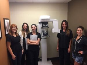
Our state-of-the-art J. Morita CBCT unit gives our doctors the confidence to treat even the most complex root systems with precision and care.
When CBCT Makes the Biggest Difference
3-D imaging is especially valuable for:
-
Retreatment cases – when a previous root canal has failed or new symptoms appear.
-
Dental trauma – to determine the extent of cracks, fractures, or internal resorption.
-
Surgical endodontics (apicoectomy) – where pinpoint accuracy is critical.
-
Complex molars or curved roots – ensuring all canals are located and properly treated.
In these scenarios, CBCT can be the difference between uncertainty and complete, confident care.
Safe, Efficient, and Focused on You
CBCT scans are safe and quick, delivering focused radiation that’s comparable to standard dental x-rays but far lower than medical CT scans. Not every patient requires 3-D imaging—but when it’s indicated, it provides invaluable information that helps protect your tooth and overall oral health.
Our Commitment to Advanced, Compassionate Care
For over two decades, Phoenix Endodontic Group has been dedicated to combining expert clinical skill with cutting-edge technology. By embracing CBCT imaging, we continue to elevate patient care—offering treatments that are more comfortable, precise, and effective than ever before.
If you’ve been referred for a root canal or are experiencing tooth pain, contact our team to learn how advanced 3-D imaging can help us diagnose and treat your tooth with accuracy and care.
Schedule your consultation today with Phoenix Endodontic Group. Experience how advanced imaging and specialized expertise can make root canal treatment more comfortable, efficient, and successful.