by Dr. Jacqueline S. Allen | Mar 15, 2018 | Blog, Endodontics, General Information, Root Canal, Technology
 Endodontists are fond of emphasizing that endodontic therapies such as root canals preserve your natural teeth, allowing you to chew, speak and eat without the downsides that come with dentures or other forms of dental restorations. Most current endodontic therapy preserves the outside of a natural tooth by placing a crown over it, while replacing the failing nerve and pulp in the canals with the latex filling gutta-percha.
However, one of the most exciting developments in professional endodontics in the past generation has been research into regenerative endodontic therapy. Instead of replacing the nerve pulp with an inert substance, this groundbreaking treatment creates and delivers healthy living tissue to replace diseased, missing or traumatized pulp.
Endodontists who are at the forefront of this research combine their knowledge of pulp biology, the proper care of dental trauma, and tissue engineering to accomplish this task. The body’s own existing cells or bioactive materials are inserted in the pulp chamber to stimulate regrowth. A related procedure, apexification, employs similar methods to grow a dentin-like substance over the apex (tip) of the tooth root, in order to improve the chances of a traditional root canal treatment succeeding when the death of the pulp in a developing adult tooth has left an open apex.
Endodontic practitioners measure the success of regenerative endodontic therapy by its ability to achieve the following treatment goals:
- Elimination of symptoms
- Increased root wall thickness and/or root length
- Positive response to pulp vitality testing
While this technique is still evolving, endodontists are following the progress of its development with great interest.
“Regenerative endodontic therapy opens the door to transforming how we approach saving natural teeth,” says Dr. Allen, an endodontist in private practice with the Phoenix Endodontic Group. “It truly may lead to a clinical situation in which we facilitate the body healing itself.”

by fisherwebdesign | Aug 10, 2017 | Blog, Cone Beam Computed Tomography, Dental Technology, Endodontics, Endodontist, Phoenix Endodontic Group, Technology
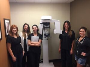 Threats to the inner pulp of your teeth can sometimes be challenging to identify and diagnose, but dental technology has come a long way in the past few years. One of the most exciting pieces of recently developed dental technology that aids endodontic specialists in their work is cone beam computed tomography, or CBCT.
Dental CBCT machines are a special type of x-ray equipment used when regular dental or facial x-rays are not sufficient. An endodontist may use this technology to produce 3-D images of teeth, soft tissues, nerve pathways and bone in a single scan. During a CBCT scan, the the machine rotates around the patient, capturing images using a cone-shaped x-ray beam. The resulting images can capture what is happening in the patient’s mouth, jaw and neck, as well as in their ears, nose and throat.
The biggest advantage of CBCT dental technology is that it allows the practitioner to visualize a patient’s condition as it actually exists in their mouth, because it is able to differentiate between many types of structures and airspaces — including bone, teeth, airway, sinuses, and soft tissue. This allows for a more accurate diagnosis and treatment planning process. CBCT can also be used after treatment to ensure that a root canal or other procedure has adequately addressed all problems that existed prior to the intervention.
Patients need to do very little to prepare for a CBCT scan, other than to wear loose clothing and leave all jewelry at home. CBCT scans are low-dose x-ray examinations compared to a standard medical CT scan.
“We’re thrilled that we can provide CBCT scans for our patients to deliver comprehensive endodontic treatment. This is a piece of dental technology that allows us to provide better care to everyone,” says Dr. Jacqueline S. Allen, who practices with the Phoenix Endodontic Group.
by fisherwebdesign | Jun 29, 2016 | Blog, Endodontics, Phoenix Endodontic Group, Root Canal, Technology
 Endodontics became a specialty in the early 1960s, but dentists have been performing root canal procedures on patients since the 1800s. Thankfully, the world of root canals has come a long way since the turn of the 19th century.
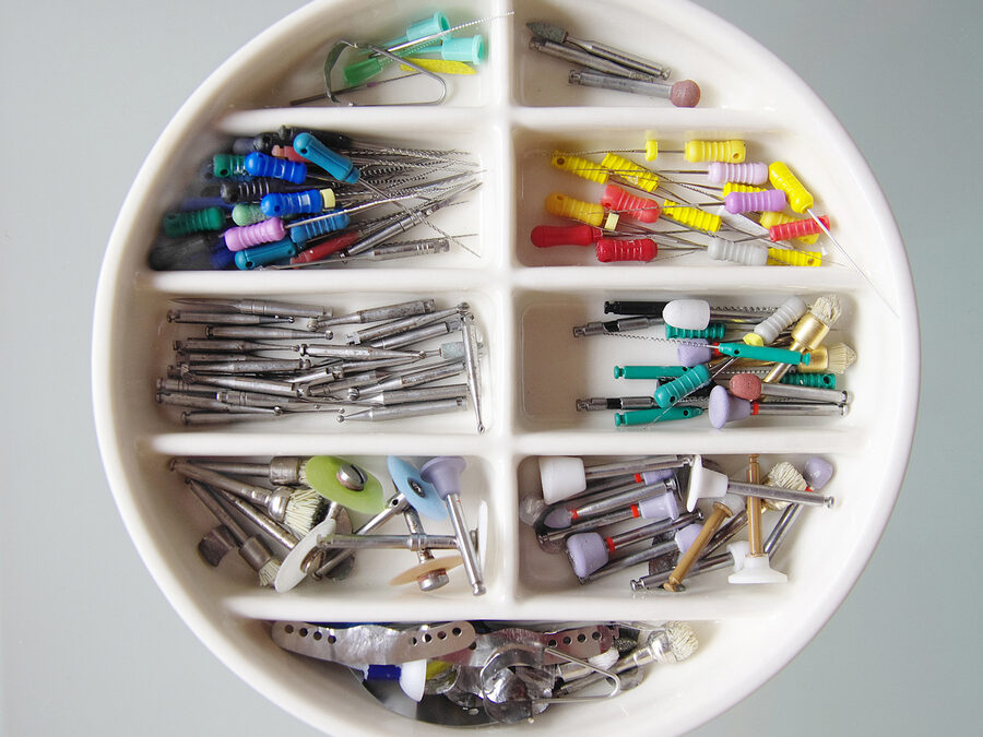
Implementing a root canal has gotten much easier, and the overall outcome of the procedure itself has improved since the introduction of nickel-titanium files. These files were first introduced in the late 1980s and are made of a unique alloy that is extremely flexible, which helps to preserve the original anatomy of the root canal. This, in turn, results in better efficiency, predictability, and improved clinical results of endodontic treatment, especially in significantly curved canals.
Digital radiography is another handy tool that we use as endodontists. Originally introduced in the 1990s, it has certainly revolutionized the field of endodontics, as well as the entire dental community, by allowing the dentist to manipulate an image and provide a much higher overall diagnostic quality. Thankfully, it does this with much less radiation than was needed to capture a standard radiographic dental film in the past.
In addition to digital radiography, the operating microscope is relatively new in the endodontic world. Magnification and fiber optic illumination are helpful in aiding the endodontist by allowing him or her to see very small details inside the teeth that that need work. Also, a tiny video camera on the operating microscope can record images of a patient’s tooth to further document the doctor’s findings.
Finally, three-dimensional radiographs (cone-beam commuted tomography) allows endodontists to help diagnose potential issues more accurately and provide treatment with unprecedented confidence. Unlike a traditional spiral CT scanner, this 3D system provides precise, crystal-clear digital images while minimizing the patient’s exposure to radiation. This system allows for unmatched visualization of anatomical detail, which aids in diagnosis, treatment planning, and the actual root canal treatment. Your doctors can use this innovative technology to quickly and easily share 3D images of the area of concern with your referring doctor, giving them an opportunity to collaborate on your care, improve your experience, and deliver a positive treatment outcome.
Although there are many other advancements in endodontics on the horizon (stem-cell regeneration of the root canal system, as well as reconstructing the teeth themselves), the current ones allow for much better diagnosis, treatment planning, and pain-free root canals than ever before.

by Dr. Jacqueline S. Allen | Jul 16, 2012 | Blog, Phoenix Endodontic Group, Technology
Technology Upgrades
Call us crazy, but here we go again. We firmly believe that all businesses should have a plan to replace technology as early as three years and no later than 5 years. The current technology in our office fits in that range and we have decided to move forward with technology upgrade project that will help us to continue to deliver the finest endodontic experience for our patients and referring doctors all under our mantra of “High Tech, High Touch.”

by Dr. Jacqueline S. Allen | Apr 23, 2012 | Blog, Endodontics, Phoenix Endodontic Group, Technology
Guest Wi-Fi
At the recent Western Regional Convention for the
Arizona Dental Association, I attended several seminars and workshops. During these learning sessions I was struck by how many participants openly checked their cellphones and I-Pads (taking notes perhaps) during the presentations. As one who has given corporate presentations to large groups of people for over 20 years, I have to say that at first I was shocked at how impolite to the presenter this behavior seemed to be.
by Dr. Jacqueline S. Allen | Apr 12, 2012 | Blog, Technology
New Scanner
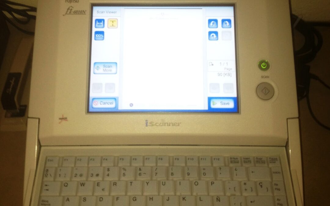
Reading the title of this post is less than exciting, huh? Well it’s exciting for me. Yesterday, we installed a new scanner in the office – a Fujitsu fi6010N SnapScan. This machine is a REAL scanner, not a copier or a printer that can do “it all” (multi-function).

 Endodontists are fond of emphasizing that endodontic therapies such as root canals preserve your natural teeth, allowing you to chew, speak and eat without the downsides that come with dentures or other forms of dental restorations. Most current endodontic therapy preserves the outside of a natural tooth by placing a crown over it, while replacing the failing nerve and pulp in the canals with the latex filling gutta-percha.


 Endodontics became a specialty in the early 1960s, but dentists have been performing root canal procedures on patients since the 1800s. Thankfully, the world of root canals has come a long way since the turn of the 19th century.


